
What are the Different Types of Microscopes?
Microscopes are essential tools used to observe and study objects or specimens that are too small to be seen with the human eye. There are many types of microscopes, all of which differ in size, style and type, designed for specific purposes and offering unique capabilities. Microscope types also vary in their physical setups, technologies, viewing results and quality levels.
As a result, choosing the right microscope for your application is important to achieve the best results. Below are a few examples of microscope types available on the market. Each type has its own advantages and applications, catering to the specific needs of various scientific fields and industries.
1. Optical Microscope
Also known as a light microscope, this is the most widely used type of microscope. These microscopes use visible light and a series of lenses to magnify and observe small specimens. The lenses are arranged between the viewer's eye or camera and the sample to focus the light and magnify the sample, making the sample easier to assess.
There are various configurations of optical microscopes:
- Compound microscopes: These microscopes work through a combination of lenses, enhancing the magnification of the sample. Under the compound microscope, the sample is first viewed as the primary image within the tube and considered again in its eyepiece.
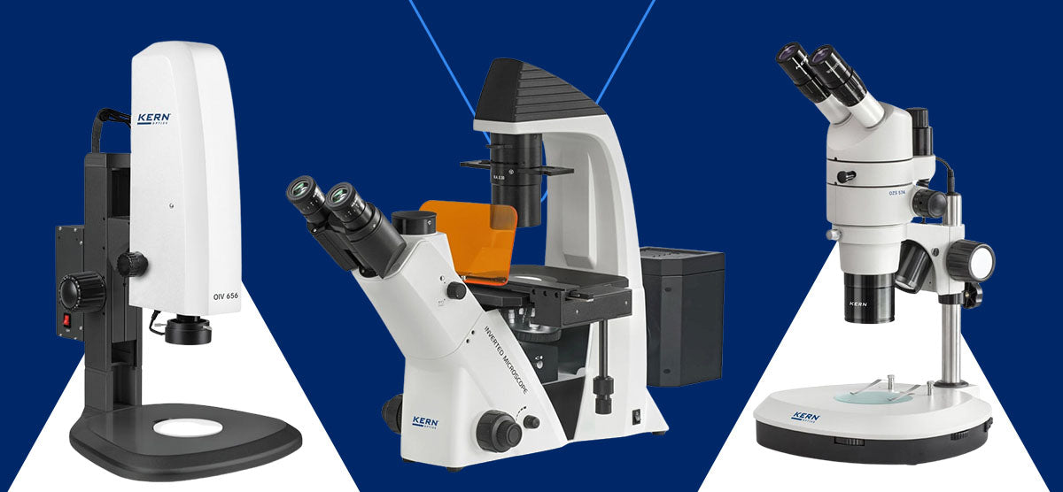
- Stereo microscopes: Also known as dissection scopes, stereo microscopes are used for dissecting microscopic specimens. They generate a three-dimensional view of objects or specimens as they reflect light from the samples. They're most suitable for solid and thick samples.
- Inverted microscopes: These microscopes are used to observe living samples on a petri dish. The user can view the liquid living cells' cultures from underneath.
2. Electron Microscope
Electron microscopes use a beam of accelerated electrons instead of light to illuminate the specimen. These microscopes apply high-voltage currents, leading to the excitation of electrons in the form of a continuous stream used as a beam of light. Electron microscopes use magnetic coil lenses to focus the electron beams to illuminate the sample.
These microscopes offer much higher magnification power and resolution compared to optical microscopes. These features make them ideal for use in biomedical research to investigate detailed cell, tissue, organelle and macromolecular complex structures.
Electron microscopes can further be divided into two types:
- Transmission electron microscope (TEM): A TEM transmits a beam of electrons through a thin specimen — as thin as 1 nanometer across — such as molecules and tissue specimens. It produces high-resolution images of the internal structure.
- Scanning electron microscope (SEM): An SEM provides detailed 3D, blue and white surface images of specimens. They're only about 10% as powerful as TEMs.
3. Scanning Probe Microscope
Scanning probe microscopes utilize a physical probe mounted on the cantilever's end to scan the surface of a specimen. This type of microscope measures various properties such as height, electrical conductivity and magnetic field. The scanning probe microscope allows you to observe highly magnified 3D specimen images in real time. These images are also probed to draw and transfer more information on the specimen.
These microscopes are often used for research in the fields of biology, chemistry and physics. Specifically, they are used in applications examining specimens occurring at nanoscale levels to determine the specimen's properties, behavior and reaction time when stimulated.
Examples include atomic force microscopes and scanning tunneling microscopes.
4. Confocal Microscope
Confocal microscopes use a laser light source and a digital camera with a pinhole. The laser scans the specimen's surface and produces 2D and 3D images on a screen without using scanning mirrors. The pinhole on the digital camera eliminates out-of-focus light, resulting in improved resolution and optical sectioning.
The goal of confocal microscopes is to eliminate issues common with fluorescence microscopes, such as allowing high-intensity UV light, which results in bleached or blurry samples. They are commonly used in biological research to study fluorescently labeled specimens.
5. Phase-Contrast Microscope
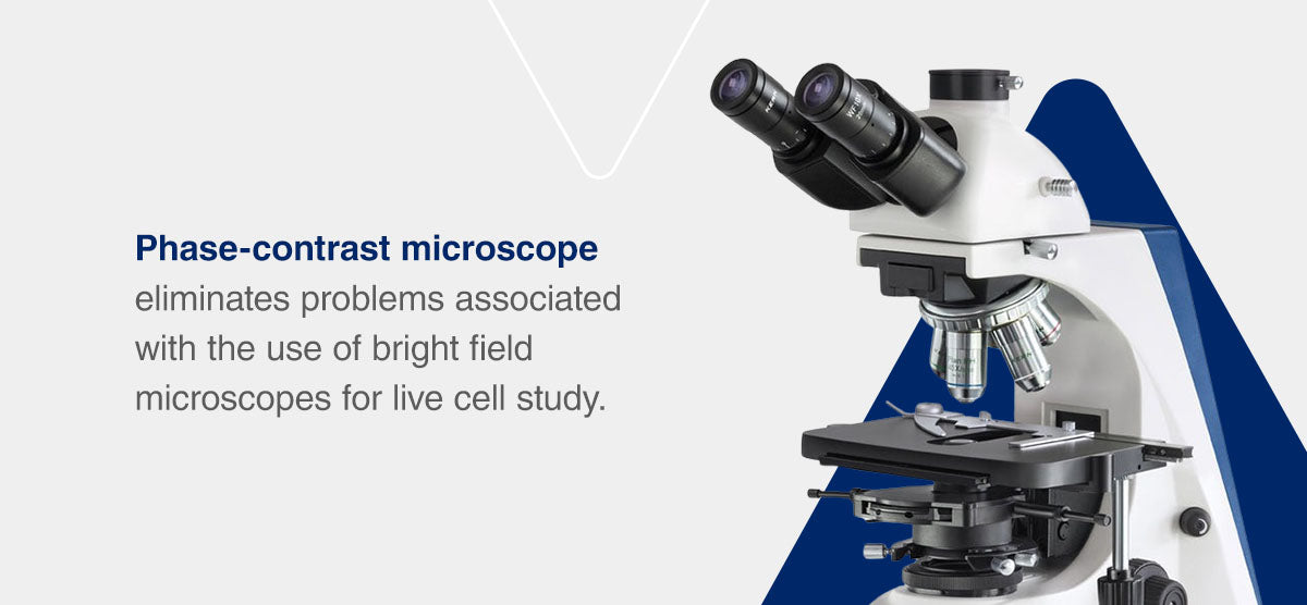
Phase-contrast microscopes enhance the contrast of transparent specimens that are difficult to see with a regular light microscope. It exploits differences in refractive index to create contrast and detail. It eliminates problems associated with the use of bright field microscopes for live cell study. Phase-contrast microscopes are ideal for looking at transparent or colorless specimens.
6. Fluorescence Microscope
Fluorescence microscopes use specific dyes or fluorescent proteins to label specific parts of a specimen. These labels emit light of a different color when excited by a particular wavelength, allowing for the visualization of specific structures or molecules. They have a powerful light source that produces high-resolution images and can be adjusted to reflect light at the desired excitation wavelength and emission.
They are utilized in many fields to look at tiny specimens like cells, microbes or fixed organic samples. Scientists prefer using fluorescence microscopes to perform life science research.
7. Polarizing Microscope
Polarizing microscopes are used to examine minerals, crystals and other anisotropic materials in fields like chemistry, geology, pharmaceuticals and petrology. They employ polarized light and specialized filters to study the optical properties and structures of these materials.
The light, which can be transmitted or reflected, is filtered using a polarizer, controlled by an analyzer and illuminated on the sample to show differences in structure, texture, color and density. The polarizer focuses the light onto a single plane while the analyzer controls the amount and direction of the light used to illuminate the sample.
Polarizing microscopes have impressive anti-reflective properties. These properties make them suitable for viewing birefringent materials — materials with two refractive indices — and isotropic materials, which have properties that remain the same when in all directions. They also have a high light sensitivity, which makes them ideal for qualitative and quantitative sample analysis.

Browse Microscopes at MSE Supplies
MSE Supplies is a reliable distributor of high-quality equipment and materials used by innovative companies and research companies. We offer a wide range of standard and custom-made microscopes to ensure we meet customer-specific requirements. We can help you source different kinds of microscopes from leading manufacturers, like Kern & Sohn and Curiosis, at the best market prices.
Contact us today to request more information about our products and services.